The Antibody Society

the official website of the antibody society

An international non-profit supporting antibody-related research and development.

  • LOG IN
  • BECOME A MEMBER
  • About
    • Mission & Activities
    • Directors and Officers
    • The Antibody Society’s Committees
      • Communication & Membership Committee
      • Meetings Committee
      • AIRR Community Working Groups & Subcommittees
    • Sponsors & Partners
  • Society meetings
    • Computational Antibody Discovery: State of the Art
      • Computational Antibody Discovery Symposium Participants
    • Harnessing Cytokines for Cancer Immunotherapy Symposium
    • Biopharmaceutical Informatics Symposium
    • Emerging Cancer Therapies Leveraging Gamma-Delta Effector T cells Symposium
    • Emerging Immunotherapeutics for Ovarian Cancer Symposium
    • AIRR Community Meetings
    • Antibody Engineering & Therapeutics (US) 2024
      • 2022 Antibody Engineering & Therapeutics
      • 2020 Antibody Engineering & Therapeutics
      • 2019 Antibody Engineering & Therapeutics
      • 2018 Antibody Engineering & Therapeutics
      • What is INN a Name?
        • INN issue updates
    • Antibody Engineering & Therapeutics Europe 2024
      • Scientific Advisors, Antibody Engineering & Therapeutics Europe
    • FOCIS Symposia
  • AIRR Community
    • AIRR News
    • AIRR Community Seminar Series
    • AIRR Publications
    • AIRR Meetings
      • AIRR Community Meeting VII – Learnings and Perspectives
      • AIRR Community Special Event 2023  – Zooming in to the Community II
      • AIRR Community Meeting VI: “Exploring New Frontiers”
      • AIRR Community Meeting V: “Zooming in to the AIRR Community”
      • AIRR Community Meeting V Pre-Meetings
        • AIRR-seq in the Pandemic
        • AIRR-seq Biological Standards and Workflows
      • AIRR Community Special Event: “Response to COVID-19”
      • AIRR Community Meeting IV: “Bridging the Gaps”
      • AIRR Community Meeting III
        • Day 1
        • Day 2
        • Day 3
        • Day 4
      • AIRR Community Meeting II
      • AIRR Community Meeting I
    • AIRR Community Working Groups
      • Biological Resources Working Group
      • Common Repository Working Group
      • Diagnostics Working Group
      • Germline Database Working Group
      • Legal and Ethics Working Group
      • Software Working Group
      • Standards Working Group
    • AIRR Community Sub-Committees
      • Communications Sub-Committee
      • Executive Sub-Committee
      • Inferred Allele Review Committee
      • Meetings Sub-Committee
      • Strategic Planning Sub-Committee
    • AIRR Data Commons
    • AIRR Community Calendar
    • AIRR Community Webinar Series
    • On AIRR – An AIRR Community Podcast
    • AIRR Community Resources
    • AIRR Community Service Prize
  • Members only
    • Login
    • Note to members
    • Member discount codes
    • 2024 Calendar of Events
    • James S. Huston Antibody Science Talent Award
      • 2022 James S. Huston Antibody Science Talent Award Recipient
      • 2021 James S. Huston Antibody Science Talent Award Recipient
      • 2020 James S. Huston Antibody Science Talent Award Recipient
      • Huston Award Criteria
    • Research Competitions
      • Research Competition Winners
    • Science Writing Competition
      • Science Writing Competition Winners
    • Imaging Competition
      • Imaging Calendar Competition winners
        • The Antibody Society 2024 Calendar
    • Antibodies in early-stage studies
    • Presentations
  • Upcoming meetings
  • Web Resources
    • Society Publications
    • Antibody News
    • Antibody therapeutics approved or in regulatory review in the EU or US
      • Antibody therapeutics product data
    • Antibodies in late-stage clinical studies
    • Research Resources
    • Education Resources
  • Career Center
    • Career Shorts
  • Learning Center
    • Upcoming Webinars
    • Snakebite antivenoms: Global challenges and progress toward recombinant antibody therapeutics
    • Adaptive Immune Receptor Repertoires
    • Antibody Discovery & Development
    • Commercializing Antibody Therapeutics
    • Antibodies to Watch
    • Antibody Validation
      • 4th International Antibody Validation Meeting, Sep 2023
You are here: Home / Archives for The Antibody Society

Join us on April 7th for our next webinar, Precision Execution of Bispecifics at Scale from Design to Delivery!

March 28, 2022 by The Antibody Society

Thursday April 7, 2022 11am ET / 5pm CET
Speaker: Dr. Lisa Prendergast, Associate Director of Expression System Sciences in Licensing at Lonza

Registration for our next webinar, “Precision Execution of Bispecifics at Scale from Design to Delivery“, is now open!

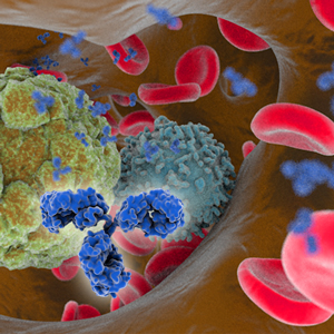
Novel therapeutic modalities such as bispecific antibodies are increasingly being explored as more effective alternatives to monoclonal antibodies for a range of diseases. Therapeutics such as bispecifics, can have a combinatorial effect by targeting two antigens,  resulting in treatments with enhanced utility, higher efficacy, fewer side effects and less resistance compared to mAbs.

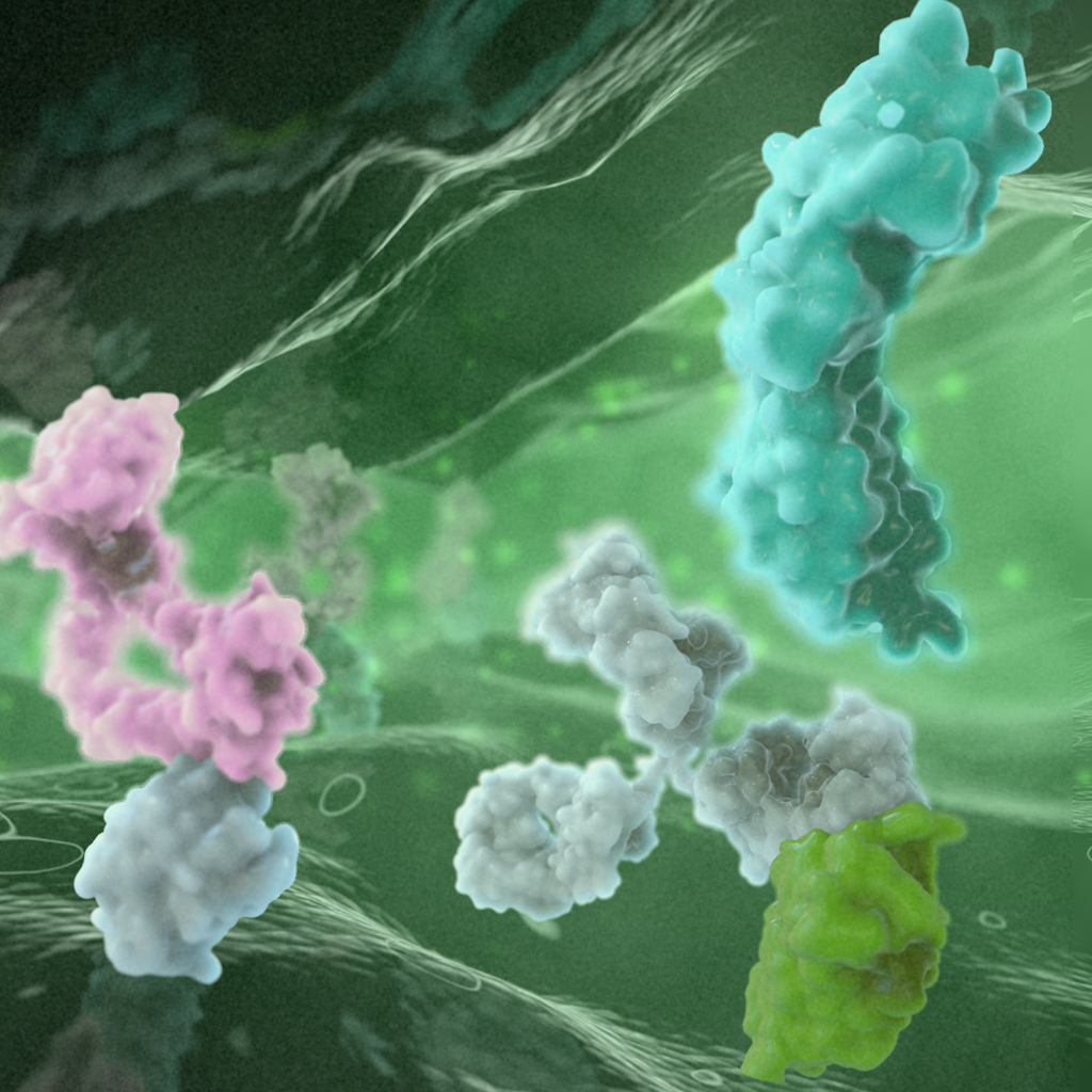
Generating a bispecific antibody, which is correctly and stably paired, is a major production concern. Many solutions require significant changes to native antibody structure, which increases antibody complexity and forces adaptation of downstream processes. While a various platforms have been developed to mitigate Heavy-Light chain (HC-LC) mispairing, there are many other rate limiting steps for efficiently expressing these molecules in a CHO system. bYlok® technology is a design engineering approach that stabilise the interaction between the HC and LC, essentially removing the mispairing problem whilst retaining a more natural antibody structure.

This presentation will introduce you to a mechanistic review of the bispecific pipeline to demonstrate how a various tools and technologies can enable you execute bispecifics.  Case studies will be presented to show how the bYlok® technology can be used to stabilise and select for novel bispecifics from a panel of parental immunotherapeutic mAbs. Our data demonstrates that correct heterodimerisation can be achieved consistently and how standard downstream purification processes can be used during production.

Register here!

Filed Under: Antibody therapeutics pipeline, Bispecific antibodies, Manufacturing Tagged With: antibody therapeutics, bispecific

Clinical-stage ROR1xCD3 bispecific antibodies with potential for broad cancer specificity

March 22, 2022 by The Antibody Society

Antibody Engineering & Therapeutics, held in December 2021, offered many opportunities to hear exciting and informative presentations by experts in the field. We are pleased to present here a summary of a lecture given in the “Immune Cell Recruitment and Redirection” session by Prof. Kerry Chester. The summary was kindly written by Dr. Czeslaw Radziejewski.

Clinical-stage ROR1xCD3 bispecific antibodies with potential for broad cancer specificity.
Kerry Chester, Professor of Molecular Medicine at University College London and CSO of Novalgen.

The leading molecule of Novalgen is NVG-111, a first-in-class tandem T-cell engager in single-chain variable fragment (scFv) format. One arm of NVG-111 targets a T-cell coreceptor, CD3, while the second binds to the tumor-associated tyrosine kinase-like receptor ROR1. ROR1 was cloned in 1992 from a neuroblastoma cell line. (1) The function of ROR1 as a tyrosine kinase is still poorly understood, although some studies show evidence of its intrinsic tyrosine kinase activity. ROR1 is a cell-surface oncofetal antigen, expressed during embryogenesis and largely absent in normal adult organs, with only low-level expression on adipocytes, pancreas, and parathyroid glands. In contrast to the lack of expression in healthy tissues, ROR1 is present in a wide range of cancers and cancer initiating stem cells. It is expressed in both hematological malignancies and in solid tumors. (2)

ROR1 has three extracellular domains: Kringle, Frizzled and Ig-like domain. ROR1 sequences of extracellular domain (ECD) are highly similar between different species. For example, there is 97.6% identity between mouse and human ROR1 ECD. Many years after the initial ROR1 discovery, its ligand was identified as Wnt-5a, one of the Wnt family signaling molecules. Unlike other ROR1 clinical candidates under development, the anti-ROR1 arm of NVG-111 binds to ROR1 Frizzled domain.

Novalgen began the development of NVG-111 by immunizing rats with recombinant extracellular domain of ROR1. The majority of the resulting antibodies bound to Ig-like domain, none bound to Kringle domain, and only one clone (clone F) bound to Frizzled domain. Clone F was selected for further development. Using flow-cytometry, Novalgen demonstrated binding of clone F to a large number of human cancer cell lines. Clone F was humanized and used to format a bispecific scFv with humanized anti-CD3. NVG-111 binds to mouse and to human ROR1 with low nanomolar affinity, but the anti-CD3 arm does not bind to mouse CD3.

In preclinical studies NVG-111 was effective in in-vitro and in an in-vivo mice model of hematological malignancies, and it demonstrated the ability to kill solid tumor in an established PANC-1 mouse xenograft model of human pancreatic carcinoma. NVG-111 also demonstrated killing in models of advanced solid tumors. It eliminated CD44+/CD24- cancer stem cells in a solid tumor model of triple-negative breast cancer. It induced dose-dependent killing in chronic lymphocytic leukemia (CLL) patient samples where patient CLL cells were cocultured with autologous T cells with EC50 in the range of 4-100 pg/ml. NVG-111 showed T cell-mediated killing of mantle cell lymphoma (MCL) cells that was as effective as killing by blinatumomab, which binds CD3 and CD19, but with 2—30% lower levels of cytokine release (measured as interferon gamma) than blinatumomab, suggesting lower risk of cytokine-release syndrome. Toxicity studies performed in mice using AAV expressing NVG-111 showed lack of toxicity at levels 20- to 1000-fold of expected steady-state levels in clinical dose. Because over 90% of CLL/MCL patients are ROR1 positive, the current focus of Novalgen clinical studies are these two hematological malignancies. Importantly, ROR1 is not expressed on normal B cells, therefore risk of B cell aplasia is expected to be reduced.

1. Masiakowski P, Carroll RD. A novel family of cell surface receptors with tyrosine kinase-like domain. J Biol Chem. 1992;267(36):26181-90.

2. Yuming Zhao et al. Tyrosine kinase ROR1 as a target for anti-cancer therapies. Front. Oncol., 11:680834. doi: 10.3389/fonc.2021.680834.

Filed Under: Antibody therapeutic, Bispecific antibodies, cancer Tagged With: antibody therapeutics, bispecific, cancer, ROR1, T-cell engager

Discovering and Targeting Neo-epitopes in Cancer

March 17, 2022 by The Antibody Society

Antibody Engineering & Therapeutics, held in December 2021, offered many opportunities to hear exciting and informative presentations by experts in the field. We are pleased to present here a summary of a plenary lecture by Prof. James Wells (USCF), kindly written by Dr. Czeslaw Radziejewski.


Discovering and Targeting Neo-epitopes in Cancer.
James Wells
, Professor and Chair, Department of Pharmaceutical Chemistry, UCSF

Professor Wells presented the plenary lecture on the identification of cancer-associated proteolytic neo-epitopes in cell membrane proteins and the identification of novel cancer-specific MHC-1 peptide complexes. Cell surface proteins are the targets of most biologic and small molecule drugs. Professor Wells and colleagues use cell surface proteomics to examine changes in the cell surface proteins upon transformation with oncogenes such as KRAS, HER2, EGFR, BRAF, MEK, and Myc. Ecto-domains of identified proteins, which generally belong to the single pass trans-membrane class, are expressed as Fc fusion proteins and antibodies are generated against these proteins via screening phage libraries. Specificities of the antibodies are verified by testing against full-length trans-membrane proteins expressed by cells transfected with appropriate vectors.

Proteolysis is a primary post-translational modification of cell surface proteins. There are approximately 500 human proteases, and proteolysis plays an important role in disease progression, such as angiogenesis, invasion and metastasis, inflammation, and immune evasion. Well’s lab is exploring methods to identify proteolytic cleavage sites on the surfaceome of cancer cells.[1] To accomplish this, they devised a technology called N-terminomics, which uses the peptide ligase called subtiligase. Subtiligase ligates peptide esters to the N-terminus of a protein or a peptide. This enzyme can be used for other purposes, such as peptide cyclization and protein bioconjugation. The lab used peptides tagged with biotin or fluorescently labelled in conjunction with mass spectrometry to identify sites of proteolytic cleavage.[2,3] Prof. Wells showed an example of this strategy used to identify sites of cleavage by caspase in the proteome of a human cell line in which apoptosis was induced. This approach, however, identified only a limited number of cleaved proteins. In the next implementation of the strategy, cells were directly transfected with subtiligase. This strategy allowed the identification of hundreds of extracellular proteins that were proteolytically modified.

The newest strategy invented in Prof. Wells’ lab (unpublished) involves tethering subtiligase to glycans of cell surface proteins instead of transacting cells. Using this latest strategy in Kras-transformed cells, 611 cell surface cleavage events were observed. In HER2-transfected cells, 267 cleavage events were observed and the majority of events were not related to cleavage of signal peptide from extracellular proteins. Interestingly, the extent of proteolytic modification of some proteins in oncogene-transformed cells can either increase or decrease. Similarly, expression levels of the same proteins also change in both directions. N-terminomics of Kras- and HER2-transformed cells was thus different.

This study also identified an interesting protein called CDCP1, which has cleavage and expression that is upregulated in pancreatic cancer. The cleavage is indeed specific to cancer cells. Three closely nested cleavage sites were found in CDCP1. Antibodies (CL03.2) were developed in the lab against the cleaved form  of CDCP1. Cells containing the cleaved form were efficiently killed by the anti-CDCP1 antibody formatted as an antibody-drug conjugate (ADC). In Jurkat cells, an anti-CD3/anti-CDCP1 bispecific single-chain variable fragment showed killing activity. For in vivo studies, mouse-specific antibodies toward the truncated form of CDCP1 were generated and used to produce an auristatin (MMAF)-based ADC. An ADC against the truncated form of CDCp1 was well tolerated in non-tumor-bearing mouse, but the animals lost weight when treated with an ADC targeting the full-length protein. In a study of mice bearing xenograph tumors, the animals were administered antibody against the truncated form that was radiolabeled with isotope Lu 177 and a dramatic decrease of tumor growth was observed.

[Read more…]

Filed Under: cancer Tagged With: Antibody drug conjugates, antibody therapeutics, bispecific, cancer

Application of Machine Learning and Informatics in Antibody and Protein Research

March 10, 2022 by The Antibody Society

Antibody Engineering & Therapeutics, held in December 2021, offered many opportunities to hear exciting and informative presentations by experts in the field. We are pleased to present here a summary of a plenary lecture by Prof. Charlotte Deane (University of Oxford), kindly written by Dr. Czeslaw Radziejewski.

Application of Machine Learning and Informatics in Antibody and Protein Research
Charlotte Deane, Professor of Structural Bioinformatics, Department of Statistics, University of Oxford

Machine learning relies heavily on the availability of large databases. Three databases for antibody research were developed in Prof. Dean’s lab: OAS (Observed Antibody Space),[1] SAbDab (Structural Antibody Database),[2] and Thera-SAbDab (database of immunotherapeutic variable domain sequences). OAS contains about 2 billion redundant antibody sequences across diverse immune states, organisms, and individuals. SAbDab is a fully automated self-updating collection of publicly available antibody structure data. It contains 5650 structures, but about 1000 truly non-redundant structures, 4213 antigen-antibody complexes and 890 structures of nanobodies. Thera-SabDob contains 696 structures as of October 2021. In addition, the lab has a CoV-AbDab database that contains sequences and structures for coronavirus antibodies for SARS-CoV-2, SARS-CoV-1 and MERS-CoV. This database contains about 5000 data points. The lab developed the SAbPred suite of tools for antibody prediction, comprising AntibodyBuilder, SPHINX, SCALOP, PEARS, ANARCI, ABangle, Hu-mAb SAAB+,TAP, Epitope Profiling SPACE and Ab-Ligaty. SCALOP, ABodybuilder, SPHINX are designed for building antibody models. ABlooper tool builds complementary-determining region (CDR) structures. ABangle is a tool for calculating and analyzing the VH-VL orientation in antibodies. TAP (Therapeutic Antibody Profiler) considers the drug-like properties of therapeutic antibodies.[3] It evaluates variable domains in antibody of interest using five developability criteria derived from post clinical Phase 1 antibody therapeutics. Epitope Profiling-SPACE and Paratyping Ab-Ligity can used to determine if two antibodies with divergent sequences can bind to the same epitope.[4] ANARCI is a tool for annotating antibody sequences and Hu-Mab is a computational tool for antibody humanization. Dlab is a deep learning method for virtual screening of antibody sequences that can bind specific antigens.

Professor Deane provided examples of using some of her computational tools. Antibody humanization is currently inefficient, as it is carried out experimentally in a largely trial and error process. Applying machine learning to an edited OAS database (with redundancies removed) led to classifiers that could distinguish between human and non-human antibody variable domain sequences. These classifiers were used to create the computational humanization tool Hu-mAb. Available sequences of therapeutic antibodies from different stages of development were subjected to Hu-mAb analysis. The high Hu-mAb scores correlated with low observed immunogenicity of an antibody and low scores correlated with higher observed immunogenicity. Twenty-five experimentally humanized antibody sequences for which rodent or rabbit precursor sequences were available were assessed by Hu-mAb. Most of the mutations that Hu-mAb generated were either the same or chemically similar for VH (77% and 85%, respectively) and for VL (59% and 58%, respectively). Hu-mAb suggested overall fewer mutations and fewer mutations to VH-VL interface than the experimental approach, therefore such humanized antibodies would more likely have preserved structure and function.

The Therapeutic Antibody Profiler evaluates properties thought to determine antibody developability, including CDRH3 or total CDR length; patches of surface hydrophobicity across CDR vicinity; patches of positive charges and negative charges across CDR vicinity; and structural Fv charge symmetry. These properties are related to aggregation, viscosity, poor expression and polyspecificity of antibody molecules.[5] TAP was applied in a study that used 137 post Phase 1 therapeutic models,14000 representative Human Antibody Models and 2 datasets of MedImmune Developability Failures. The study revealed that therapeutic antibodies tend to have shorter CDRH3 and smaller hydrophobic patches than natural ones. However, positive and negative patches of natural and therapeutic antibodies have similar profiles and Fv charge symmetry is also very similar. Both therapeutic and natural antibodies have an aversion to strongly oppositely charged VH and VL chains.

ABlooper [6] uses similar architecture as AlphaFold. It predicts structures of all six CDR loops and estimates the accuracy of prediction. The root-mean-square deviation from AlphaFold2 for CDRH3 prediction (2.87A) were comparable with ABlooper (2.49 A). Unlike AlphaFold2, ABlooper generates a series of predicted structures from which a prediction of accuracy can be estimated. If the predicted structures are widely divergent, then the quality of prediction is low. ABlooper is also much faster than other deep learning methods such as AlphaFold (100 structures predicted in 5 second vs one structure in 20 min). All tools are available freely for academic institutions.

  1. Olsen TH, Boyles F, Deane CM. Observed Antibody Space: A diverse database of cleaned, annotated, and translated unpaired and paired antibody sequences. Protein Sci. 2022 Jan;31(1):141-146. doi: 10.1002/pro.4205.
  2. Schneider C, Raybould MIJ, Deane CM. SAbDab in the age of biotherapeutics: updates including SAbDab-nano, the nanobody structure tracker. Nucleic Acids Res. 2022 Jan 7;50(D1):D1368-D1372. doi: 10.1093/nar/gkab1050.
  3. Raybould MIJ, Deane CM. The Therapeutic Antibody Profiler for Computational Developability Assessment. Methods Mol Biol. 2022;2313:115-125. doi: 10.1007/978-1-0716-1450-1_5.
  4. Wong et al. Ab-Ligity: identifying sequence-dissimilar antibodies that bind to the same epitope. MAbs 2021. DOI: 10.1080/19420862.2021.1873478.
  5. Khetan et al. Current advances in biopharmaceutical informatics: guidelines, impact and challenges in the computational developability assessment of antibody therapeutics. MAbs 2022. DOI: 10.1080/19420862.2021.2020082.
  6. Abanades B, Georges G, Bujotzek A, Deane CM. ABlooper: Fast accurate antibody CDR loop structure prediction with accuracy estimation. Bioinformatics. 2022 Jan 31:btac016. doi: 10.1093/bioinformatics/btac016.

Filed Under: Bioinformatics Tagged With: bioinformatics, machine learning

Untangling Pandemics in a Data-Driven World. The Evolution of SARS-CoV-2

March 4, 2022 by The Antibody Society

Antibody Engineering & Therapeutics, held in December 2021, offered many opportunities to hear exciting and informative presentations by experts in the field. We are pleased to present here a summary of a plenary lecture by Prof. Kristian Andersen (Scripps Research Institute), kindly written by Dr. Czeslaw Radziejewski.


Untangling Pandemics in a Data-Driven World. The Evolution of SARS-CoV-2.
Kristian Andersen, Professor of Immunology and Microbiology, Scripps Research Institute

Professor Andersen’s lab conducts genomic epidemiology of different viruses using miniaturized PCR testing and large-scale genomic sequencing. Previously, his lab studied Lassa, West Nile, Ebola, and Zika viruses, and now the lab is examining SARS-CoV-2. The goal of the research to understand the emergence of new viruses and their local transmission,[1] as well as the evolution and spread of these viruses. In order to understand connectedness of the sequences, samples of virus are taken from the infected populations, then viruses are sequenced and analyzed.

SARS-CoV-2 was first detected in December 2019 in people who frequented the Wuhan Huanan market in Guangdong province, China. Based on large-scale sequencing, it was possible to estimate that the epidemic started in November 2019. SARS-CoV-2 is remarkably similar to SARS-CoV-1 in terms of the receptor in humans (ACE2), the animal reservoir (bats), and the fact that both are associated with wet markets.[2] The number of introductions in humans is at present unknown and any intermediate hosts are still also unknown. For SARS-Cov-1 the intermediate hosts were civets and raccoon dogs. Curiously, both viruses were introduced into humans in the month of November, SARS-Cov-1 in 2002, SARS-Cov-2 in 2019. Since 2002, trade and farming of wild animals have decreased in China, so the risk of multiple spillover was reduced. A farm in Hubei was previously found to have animals infected with SARS-CoV-1. Viral sequences determined in animals were very similar to those found in humans infected with SARS-Cov-1. The same farm recently had civets infected with SARS-CoV-2.

Since 1965, nine different coronaviruses have emerged in human population. Coronaviruses are part of Sarbecoviruses, which are widespread in Southeast Asia. Horseshoe bats are also widespread in that region and they are a reservoir of Sarbecoviruses. SARS-CoV-2 spreads easily and, unlike a seasonal influenza, it can infect both upper and lower parts of the respiratory system as well as other organs. High infectivity of many variants of CoV-2 can be attributed to the presence of the receptor binding domain (RBD) and also the presence of a polybasic cleavage site that allows the spike protein to be processed into two subunits and facilitates fast and widespread infection. The SARS-CoV-2 RBD is similar to the RBD of other viruses that infect animals such as bats and pangolins. SARS CoV-2 shows a very high rate of evolution, which in the past two years has resulted in multiple variants.[3] New variants have rapidly displaced the old ones. Omicron, which presumably emerged as a result of immune escape, has an extremely mutated lineage. For example, it has 40 mutations in the spike protein compared to original virus, most of which appears on the outside RBD of the spike protein. The mutations may lead to further optimization of binding to AC2, but also potentially usage of a coreceptor is involved. The spread of omicron is much faster than other variants. This variant is cable of causing new infection and reinfection. Almost entire the human population is susceptible to omicron infection, and SARS-CoV-2 now appears to be endemic.

In summary, SARS-Cov-2 is a fast-evolving virus and several factors such as evolutionary rate, mutational supply, mutational tolerance will determine its further evolution. However, we now have tools to fight the pandemics, including vaccines, anti-viral drugs, rapid tests, masks and better understanding of the virus.

1. Zeller et al. Emergence of an Early SARS-CoV-2 Epidemic in the United States. Cell 184(19):4939-4952, 2021. DOI: 10.1016/j.cell.2021.07.030.

2. Holmes et al. The Origins of SARS-CoV-2: A Critical Review. Cell 184(19):4848-4856, 2021. DOI: 10.1016/j.cell.2021.08.017.

3. Washington et al. Emergence and Rapid Transmission of SARS-CoV-2 B.1.1.7 in the United States. Cell 184(10):2587-2594, 2021. DOI: 10.1016/j.cell.2021.03.052.

Filed Under: COVID-19, SARS-CoV-2 Tagged With: COVID-19, SARS-CoV-2

« Previous Page
Next Page »

mabs

mabs

The Official Journal of The Antibody Society

Career Center

Our Career Center is a premier resource to connect highly qualified talent with matching career opportunities. Visit for details on over 800 jobs!

AIRR Community

AIRR Community

The Adaptive Immune Receptor Repertoire Community is a research-driven group organizing around the use of high-throughput sequencing technologies to study antibody/B-cell and T-cell receptor repertoires.

Recent Posts

  • World Cancer Day 2024 – Antibody therapeutics for cancer indications February 5, 2024
  • The Antibody Society is hiring! January 23, 2024
  • The Antibody Society announces the election of new Directors and Officers January 19, 2024

Archives

Follow us online

  • Email
  • LinkedIn
  • Twitter
  • YouTube
  • Home
  • Privacy & Terms of Use
  • About
  • Directors and Officers
  • Advisors
  • Sponsors & Partners
  • Mission & Activities
  • Join the Society
  • Membership Levels
  • Members only
  • Login
  • Antibody therapeutics approved or in regulatory review in the EU or US
  • Meeting reports
  • Presentations
  • Contact

©2015 - scicomvisuals